
美国的研究人员National Eye Institute(NEI) have developed a way of 3D bioprinting eye tissues using patient stem cells.
Using their approach, in which three different types of immature choroidal cells are printed onto a biodegradable scaffold, the scientists say it could be possible to create an unlimited supply of patient-derived tissues. These, in turn, have the potential to help doctors better understand the mechanisms behind common degenerative retinal diseases, such as age-related macular degeneration (AMD).
“Our collaborative efforts have resulted in very relevant retina tissue models of degenerative eye diseases,” said Marc Ferrer, Ph.D., Director of the 3D Tissue Bioprinting Laboratory at NIH’s National Center for Advancing Translational Sciences. “Such tissue models have many potential uses in translational applications, including therapeutics development.”
确定解决AMD的新方法
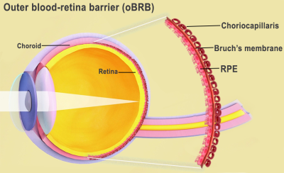
本质上是一种疾病,模糊了病人的中央vision, AMD is caused by various factors, including age, genetics, blood pressure, and dietary issues. These triggers are understood to cause lipoprotein deposits called drusen to form outside Bruch’s membrane, an extracellular matrix (ECM) located between the retina and the choroidal capillaries of the eye.
This phenomenon not only prevents the membrane from regulating nutrients and waste around the eye, but over time, causes the retinal pigment epithelium (RPE) in its outer blood-retina barrier to break down, leading to vision loss. However, while the triggers of AMD are well-known, the way it advances remains something of a mystery.
“We know that AMD starts in the outer blood-retina barrier,” explained Kapil Bharti, Ph.D., who heads the NEI Section on Ocular and Stem Cell Translational Research. “However, mechanisms of AMD initiation and progression to advanced dry and wet stages remain poorly understood due to the lack of physiologically relevant human models.”
NEI的3D生物打印计划
为了促进对AMD进展的进一步研究,Bharti及其团队提出了一种来自患者干细胞的3D生物打印组织的方法。在这种技术中,周围细胞和内皮细胞,毛细血管和成纤维细胞的关键成分(赋予组织结构)被合并为新型水凝胶。然后将该凝胶印在能够支撑细胞生长的脚手架上。
Within days of creating prototype biodegradable scaffolds, the scientists found that implanted cells had begun to mature into a dense capillary network. On day nine, the researchers decided to seed retinal pigment epithelial cells on the flip side of the scaffold, and by day 42, they found that tissues had reached “full maturity.”
Subsequent tissue analyses and genetic and functional testing showed that the resulting tissues looked and behaved similarly to a native outer blood-retina barrier. Under induced stress, they even exhibited patterns of drusen deposits underneath the RPE and progression to late dry-stage AMD, whereas the administration of Anti-VEGF drugs suppressed vessel overgrowth and restored tissue morphology.
证明了他们方法的功效,Bharti and Co。现在正在使用其生物打印的血元素屏障模型继续研究AMD。向前迈进,团队计划尝试在其过程中添加其他细胞类型(例如免疫细胞),以创建更多栩栩如生的组织,使他们能够进一步发现。
“通过印刷细胞,我们促进exchange of cellular cues that are necessary for normal outer blood-retina barrier anatomy,” added Bharti. “For example, the presence of RPE cells induces gene expression changes in fibroblasts that contribute to the formation of Bruch’s membrane – something that was suggested many years ago but wasn’t proven until our model.”

3D printing’s eye treatment potential
While the 3D bioprinting of eye tissues remains at an experimental stage, conventional 3D printing has been deployed in the production of various other optical medical procedures. In 2021, surgeons at the IsraeliGalilee Medical Center(GMC) developed a means oftreating eye socket fractures with 3D printing和增强现实(AR)眼镜。
那年晚些时候,一个团队巴塞尔大学also unveiled porous3D printed implants for treating eye socket fractures。使用a创建Prusa i3 3D打印机and PEEK filament, the team’s grafts were said to be capable of overcoming the bioinertness of their material thanks to their porous features, which could be tailored to enhance cellular repair.
在其他地方,如果无法保存患者的眼睛,医生在Moorfields眼科医院3D printed a prosthetic eye for a London man需要。使用该技术,该团队能够与传统的丙烯酸假体更现实的外观创造人造眼,该丙烯酸假体可以在短时间内制造。
The researchers’ findings are detailed in their paper titled “Bioprinted 3D outer retina barrier uncovers RPE-dependent choroidal phenotype in advanced macular degeneration。”
The study was co-authored by Min Jae Song, Russ Quinn, Eric Nguyen, Christopher Hampton, Ruchi Sharma, Tea Soon Park, Céline Koster, Ty Voss, Carlos Tristan, Claire Weber, Anju Singh, Roba Dejene, Devika Bose, Yu-Chi Chen, Paige Derr, Kristy Derr, Sam Michael, Francesca Barone, Guibin Chen, Manfred Boehm, Arvydas Maminishkis, Ilyas Singec, Marc Ferrer and Kapil Bharti.
To stay up to date with the latest 3D printing news, don’t forget to subscribe to the3D打印行业newsletter或跟随我们推特或喜欢我们的页面Facebook。
当您在这里时,为什么不订阅我们Youtube渠道?包括讨论,汇报,视频短裤和网络研讨会重播。
Are you looking for a job in the additive manufacturing industry? Visit3D打印作业在行业中选择一系列角色。
特色图像显示了人眼内部的图。通过国家眼科研究所的图像。