
Researchers in Korea and Hong Kong have used a modified 3D cell printing technique to fabricate a biomimetic blood vessel that was successfully implanted in a living rat.
According to the researchers, their approach to tissue-engineered biomimetic blood vessels outlined in their study provides a promising route for the construction of durable small-diameter vascular grafts. These 3D cell printed vascular grafts, which are used to redirect blood flow from one area of the body to another, have the potential to be used in future treatments of cardiovascular diseases.
“The artificial blood vessel is an essential tool to save patients suffering from cardiovascular disease,” comments Ge Gao, an author on the paper. “There are products in clinical use made from polymers, but they don’t have living cells and vascular functions.”
“We wanted to tissue-engineer a living, functional blood vessel graft.”
Creating functional vascular grafts using triple-coaxial cell printing
研究人员通过解释了心血管疾病是全球死亡率的主要原因之一,需要超过一百万的血管旁路/替代品surgeries annually in the United States alone.
组织工程, according to the authors of the paper, has emerged as a promising approach for creating viable small-diameter vascular grafts, which can be used to treat cardiovascular diseases through vessel implantation. However, thus far the construction of these vascular substitutes with both endothelial and muscular tissues has not been demonstrated.
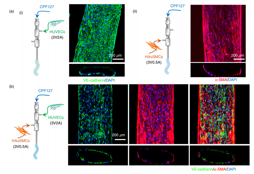
内皮细胞和平滑肌组织的定位对于移植物起作用至关重要。正如论文的作者所解释的那样,他们说:“要产生具有常规血管功能的移植物,必须实现两种关键成分的重建,即:(1)汇合和静止的内皮,提供非触及性界面,以抑制肌瘤,(以及((2)收缩的平滑肌组织可以承受血液动力学应激,表现出生理依从性并通过收缩和放松来适应局部血压的变化。”

To overcome this hurdle, the researchers identified 3D cell printing due to its ability to construct tissue/organ equivalents. The coaxial-extrusion technique in particular, which uses a special nozzle to feed one type of material into another as it is extruded, enables the creation of vessel-like structures.
为了成功地通过功能性组织工程的血管进行细胞打印,研究团队必须选择有利于细胞的生物学,既可以促进细胞功能,又可以促进细胞功能并能够直接制造血管。因此,他们开发了一种由人主动脉和脐静脉内皮细胞的平滑肌细胞制成的血管组织特异性生物学。
结合ning this bioink formulation with a triple-coaxial cell printing (RTCCP) technique thus enabled the researchers to create a functional blood vessel with a dual-layer architecture that outperforms existing engineered tissue: “The developed triple-coaxial-cell-printing technique enabled the direct construction of vascular substitutes that contain both the endothelial and muscular layer,” explain the researchers.
将这些血管作为腹主动脉嫁接成六只大鼠,并在数周内进行监测。科学家观察到了一种转化,其中大鼠的成纤维细胞在植入物表面形成了一层结缔组织,从而使制造的血管移植物作为现有生物组织的一部分集成。作者结束了这篇论文的结论:“这些发现表明,使用提出的技术构建的血管等效物是组织工程小直径血管移植物的有前途的选择。”
Moving forward, the researchers plan to continue developing their process leveraging the RTCCP technique alongside the vascular tissue-specific bioinks, specifically to increase the strength of the blood vessels closer to that of human coronary arteries. They also aim to perform long-term evaluation of vascular grafts, in order to determine what happens as they continue to develop in place and become real tissue in the implanted environment.

组织工程研究中的3D生物打印
组织工程is a rapidly evolving field, and the latest advances in 3D printing and bioprinting has enabled the technology’s application in research projects seeking to build living tissue constructs in the shape of human organs.
For example, in late 2019, researchers fromHarvard UniversitysWyss Institute开发了一种新颖的牺牲墨水写作技术,称为swift(牺牲写入功能组织)3D打印大的血管化人体器官构建块(OBBs). The researchers demonstrated the method by creating cardiac tissue that fuses and beats synchronously over a 7-day period.
大约在同一时间,研究人员Lehigh Universityin Pennsylvania also presented a new 3D printing platform that facilitated theregeneration of multiple tissuesusing spatially functionalized scaffolds. The study details how 3D printing highly organized biomaterial scaffolds can be used to regenerate two different tissues.
Just recently, in February 2020, we reported on a study from New Jersey-basedRutgers University工程师开发了一种由活细胞制成的新的适应性生物墨水3D print scaffolds for human tissue growth.
The study discussed in this article, “Tissue-engineering of vascular grafts containing endothelium and smooth-muscle using triple-coaxial cell printing,” is authored by Ge Gao, Hyeok Kim, Byoung Soo Kim, Jeong Sik Kong, Jae Yeon Lee, Bong Woo Park, Su Hun Chae, Jisoo Kim, Kiwon Ban, Jinah Jang, Hun-Jun Park and Dong-Woo Cho. The research was supported by grants funded by the Korean government, the Ministry of Trade, Industry & Energy (MOTIE, Korea), and the Ministry of Science, ICT and Future Planning. It is published inApplied Physics Reviews.
The nominations for the2020 3D印刷行业奖are now open. Who do you think should make the shortlists for this year’s show? Have your say now.
Subscribe to the3D打印行业newsletterfor the latest news in additive manufacturing. You can also stay connected by following us onTwitter和喜欢我们Facebook.
Looking for a career in additive manufacturing? Visit3D Printing Jobsfor a selection of roles in the industry.
特色图像显示,亚洲的研究人员使用三片融型细胞打印技术来构建仿生组织工程的血管,这些血管通过大鼠模型中的腹部主动脉间移植物在体内进行了评估。图像通过应用物理评论。betway必威手机版登录